
Written by: Millie Castellanos, B.S., R.T.(R)(M)(ARRT)(CRT)
Ask most patients what a mammogram feels like, and you’ll hear the same words: squeeze, flatten, pancake. (Ouch!) What these patients are actually describing is compression. For many, it's the part of the exam they anticipate the most, and often the part they fear the most. As mammographers, we understand that compression isn't about causing pain or discomfort to our patients. It's one of the most important components of producing a high-quality mammogram and it plays a vital role in the early detection of breast cancer.Why Compression Matters
Compression is much more than simply flattening and squeezing the breast. It directly impacts image quality, radiation dose, and ultimately, our ability to detect cancer.
When appropriate compression is applied, the breast tissue is spread out, reducing the overlap of normal structures that can hide abnormalities. Better tissue separation allows subtle masses, architectural distortions, and tiny clusters of calcifications to become more visible.
Without adequate and proper compression, important findings can remain hidden.
ACR Standards
According to the 1999 ACR Manual, ideal compression should be based upon these two factors:
- The maximum amount an individual patient's breast can actually be compressed.
- The amount of compression that the patient can tolerate during the exam.
The ACR manual also states that the breast should be compressed until the tissue is taut. “Taut” means that the breast should be stretched or pulled tight, with no slack. Gentle tapping of the breast should not indent the skin when the breast tissue is taut.
Measuring Compression
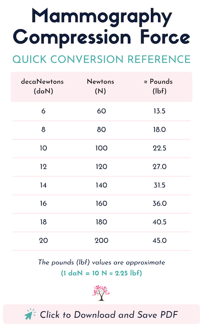
For most mammography units, compression is measured in decaNewtons, Newtons or pounds.
Under-compression can occur when insufficient force is applied to the breast. This can result in a reduction in image quality where overlapping tissue can obscure small lesions. It can also create difficulty in identifying microcalcifications due to motion. The thicker the breast tissue, when not adequately compressed, will also result in a higher dose of radiation to the patient. Repeat rates can also increase as overlapping tissue can mimic abnormalities. Inadequate or under-compression can overall lead to missed breast cancers!
Over-compression occurs when there is an excessive force applied to the breast. This can possibly cause tissue distortion. It can also increase pain and anxiety for the patient, which can result in the patient not wanting to return for her subsequent screening mammograms.
Rather than aiming for a specific compression force, mammographers should focus on effective compression - meaning compressing until the breast is adequately immobilized or taut and tissue separation has been achieved. All of this should be considered while remaining attentive to the patient’s pain tolerance.
Consistency in Compression
Applying compression consistently helps create reproducible images from one examination to the next, allowing for more reliable comparisons and potentially improving the detection of abnormalities. Making consistent compression a standard of care supports both image quality and patient outcomes.
Technologists should check their compression data, which is available on the mammography unit. Compression should be within 10% of the measurement side-to-side, with a higher compression reading (approximately 20% higher) on MLO views (Huppe et al., 2017).
The Benefits of Proper Compression
1. Improves Image Quality — Proper compression reduces tissue overlap, making breast structures easier to evaluate.
This allows radiologists to:
Simply put, better compression leads to better images.
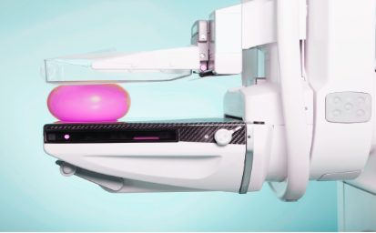
2. Reduces Motion Blur — Even the slightest patient movement during an exposure can cause blurriness and reduce image sharpness.
Proper compression stabilizes the breast, minimizing motion and producing clearer, sharper images that are easier to interpret.
3. Lowers Radiation Dose — An adequately compressed breast is thinner, allowing the x-ray beam to penetrate more efficiently.
This means:
Compression is one of the simplest ways to optimize image quality while keeping radiation doses as low as reasonably achievable (ALARA).
4. Decreases Scatter Radiation — When the breasts are properly compressed, thinner breast tissue will produce less scatter radiation.
Less scatter radiation results in:
5. Reduces Repeat Images — As mentioned above, inadequate compression may lead to:
Every repeat image increases patient anxiety and exposes the patient to additional radiation. Proper compression helps reduce any unnecessary repeat imaging.
Compression Is Not One-Size-Fits-All
As we all know, every breast is different in size, shape and density. Other factors such as, surgical history, pain tolerance, and patient anxiety can also contribute to how much compression should be applied.
Compression should always be individualized.
Communication Makes All the Difference

Patients are far more likely to tolerate compression when they understand why it's necessary. Simple explanations can ease anxiety: "Compression helps spread the breast tissue so we can see everything clearly. It also helps reduce your radiation dose and keeps the breast still so we don't have to repeat the image."
Taking just a few extra seconds to explain the purpose of compression can ease the patient’s anxiety and significantly improve the overall patient experience. Proper compression should be taut enough to produce optimal images while maintaining open communication and continuously assessing patient comfort. The goal isn't to cause pain, it's to achieve the best possible images with the least amount of discomfort to the patient.
Compressing With Purpose
Compression is one of the most powerful tools a mammographer has. Done right, alongside standardized positioning, it sharpens image quality, cuts radiation dose, reduces motion, and helps catch cancer earlier.
Every patient deserves a mammogram that gives the radiologist the best shot at finding cancer early. Pair technical skill with empathy and clear communication, and you turn a feared experience into one built on trust.
Compression isn't just about squeezing the breasts. It's about quality diagnostic images, earlier detection, and potentially, saving a life.


References:
ACR. (1999). 1999 ACR Mammography Quality Control Manual (pp. 35-36).
FDA article underscores benefits of adequate breast compression for mammography image quality. (2018, June). Imaging Technology News. https://www.itnonline.com/content/fda-article-underscores-benefits-adequate-breast-compression-mammography-image-quality
Huppe, A. I., Overman, K. L., Gatewood, J. B., Hill, J. D., Miller, L. C., & Inciardi, M. F. (2017). Mammography positioning standards in the digital era: Is the status quo acceptable? AJR. American Journal of Roentgenology, 209(6), 1419–1425.
https://doi.org/10.2214/AJR.16.17522
Miller, L., & Terry, L. (2020). Image Quality and Positioning Problem-Solving for Breast Imagers. Mammography Educators.
Roberts, C., & Marshall, J. (2019). Optimal Breast Compression in Mammography.
https://www.lunit.io/wp-content/uploads/2026/04/Optimal-Breast-Compression-in-Mammography.pdf